14
9982-1000357
9982-1000358
9982-1000349
9982-1000359
9982-1000360
3 B S c i e n t i f i c ® B i o l o g y
Anatomi ca l Mode l Human
Ske l eton & Bones
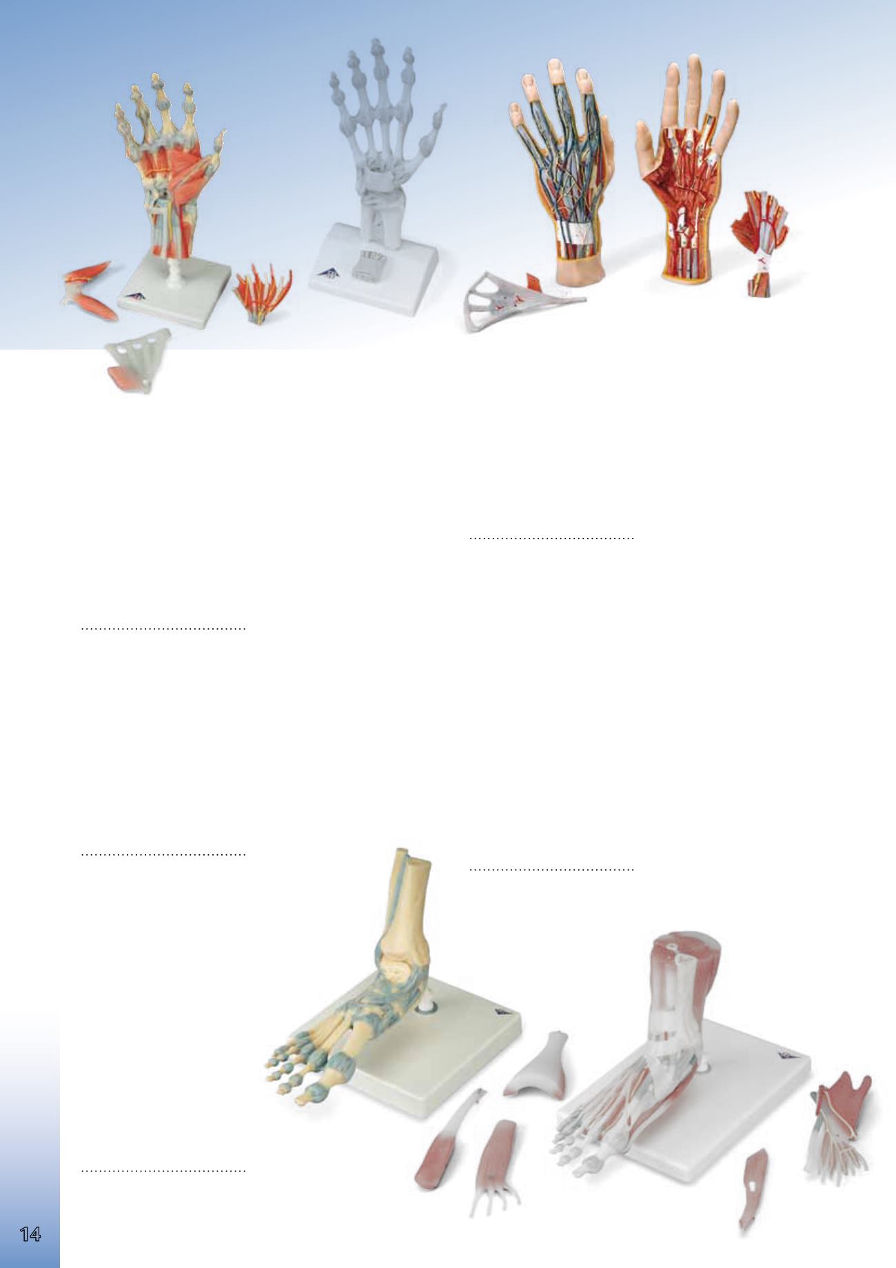
Internal Hand Structure Model, 3-part
Full size hand model showing the superficial and internal structures of
the hand, including bones, muscle, tendons, ligaments, nerves, and arter
ies (superficial and deep palmar arches). The palmar aponeurosis and plate
of the superficial tendons are removable.
28.5x13x6.5 cm; 1.2 kg
L/D/E/S/F/P/I/J www.
9982-1000349
Hand Skeleton Model with Ligaments and Carpal Tunnel
This 3 part hand model shows the anatomical details of the ligaments and
tendons found in the hand, wrist, and lower forearm. The interosseous
membrane between the radius and ulna is shown along with the bones of
the hand.The flexor retunaculum is removable and in addition there is a
removable portion that can be fitted onto the back of the model. This por
tion features the clinically important structures of the carpal tunnel such
as the flexor retinaculum, mediane nerve, and tendons.
30x14x10 cm, 0.3 kg
L/D/E/F/I/S/P/J/R/C www.
9982-1000357
Hand Skeleton Model with Ligaments and Muscles
The bones, muscles, tendons, ligaments, nerves, arteries, and veins are all
featured in this high quality 4 part model of the hand and lower forearm.
The dorsal side shows the extensor muscles as well as portions of the ten
dons at the wrist as they pass under the extensor retunaculum. The palmar
face of the hand is represented in three layers, the first two removable to
allow detailed study of the deeper anatomical layer. In additon clinically
important structures such as the median nerve and superficial palmar arte
rial arch can be examined in detail. The deepest anatomical layer allows
for study of the intrinsic muscles and deep palmar arterial arch in addition
to other details.
33x12x12 cm, 0.4 kg
L/D/E/F/I/S/P/J/R/C www.
9982-1000358
Foot Skeleton Model
with Ligaments
This detailed model displays nu
merous important ligaments and
tendons including the achilles and
peroneus longus tendons of the
ankle. The model consists of the
bones of the foot and lower por
tions of the tibia and fibula, in
cluding the introsseous membrane
found between them. All the ana
tomically important ligaments and
tendons are shown.
23x18x30 cm, 0.6 kg
L/D/E/F/I/S/P/J/R/C www.
9982-1000359
Foot Skeleton Model with Ligaments and Muscles
This model is the best of its kind for quality and value. This anatomically
detailed model of the foot and lower leg can be disassembled into 6 re
movable parts for detailed study. The model features not only the bones
but also muscles, tendons, ligaments, nerves, arteries, and veins. The fron
tal view features the extensor muscles of the lower leg. The tendons can be
followed on their passage under the transverse and crucial crural ligaments
all the way to their insertion points. In addition all tendon sheaths are visi
ble. On the dorsal portion of the model the gastrocnemius muscle is re
movable to reveal deeper anatomical elements. The sole of the foot is
represented in three layers; the first layer displaying the flexor digitorum
brevis. This muscle can be removed revealing the quadratus plantae, the
tendon of the flexor digitorum longus, and the flexor hallucis muscle.
This second layer is in turn removable to display even deeper anatomical
details.
23x26x19 cm, 1.1kg
L/D/E/F/I/S/P/J/R/C www.
9982-1000360


