60
9982-1005129
9982-1002504
9982-1002505
9982-1005130
9982-1005131
9982-1000523
9982-1000524
9982-1005124
9982-1002506
3 B S c i e n t i f i c ® B i o l o g y
Botani ca l Mode l s / Ce l lul ar Biology Mode l s
Pl ant Ce l l / Anima l Ce l l
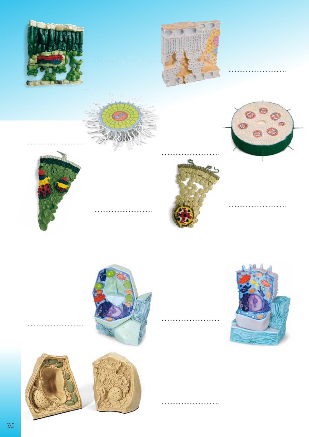
Block Model of Leaf Sructure
Cube-shaped detail of the pedate,
bifacial deciduous leaf of the
Christmas Rose (Helleborus niger)
enlarged by a factor of 1500, with
stoma on the underside.
30x30x9cm; 1.4kg
&
L/D/E/F/I/S/P/J/R/C
9982-1002504
Dicotyledons –
Stem Cross-Section
Cross-section of a Creeping Butter-
cup stem with collateral open vas-
cular bundles. The model shows
the typical stem structure of a dico
tyledon enlarged by a factor of 250.
28x7cm; 0.8kg
&
L/D/E/F/I/S/P/J/R/C
9982-1002506
Relief Model of Leaf Structure
Representation of the histological
structure of a leaf (Ligustrum),
magnified 500 times.
6.5x24x26 cm; 1.4 kg
&
E
9982-1005129
Absorption Zone of the Root
With the example of the white
mustard (sinapis alba) this relief
model shows the absorption
zone of a dicotyledonous plant.
43x43x8 cm; 1.5 kg
&
E/D/H
9982-1002505
Tissue Structure of the
Sunflower Stem
(Helianthus annuus)
Detailed longitudinal and lateral
view 200 times magnified.
&
E
9982-1005130
Tissue Structure of the Butter-
cup Root (Ranunculus)
Longitudinal and lateral view at
400 times magnification.
&
E
9982-1005131
The Plant Cell, magnified 500,000-1,000,000 times
The two-piece model presents the structure of a typical plant cell with
cytoplasm and cell organelles, as viewed from an electron microscope.
For better illustration, all important organelles are raised and displayed
in colour, e.g.:
• Cell wall
• Cell membrane
• Nucleus
• Smooth Endoplasmic Reticulum
• Rough Endoplasmic Reticulum
• Ribosomes
• Chloroplasts
• Mitochondria
• Dictyosomes/Golgi apparatus
20x14x32 cm; 0.8 kg
&
E/D/S/F/P/I/J
9982-1000524
Comparison Models Animal and Plant Cell
These enlarged models of an animal cell and a plant cell enable visual
teaching about their structures, as well as their similarities and differences.
The cell structures are numbered and identified, and the product manual
also includes reproducible illustrations for use in testing. Furthermore, the
set contains 12 electron microscopic illustrations of different cell structures.
Supplied with teacher’s notes in English.
16x15x9 cm; 1 kg
&
E
9982-1005124
The Animal Cell
The two-piece model shows the form and structure of a typical animal
cell as viewed from an electron microscope. For better illustration, all
important organelles are raised and displayed in colour, e.g.:
• Nucleus
• Mitochondrion
• Smooth Endoplasmic Reticulum (ER)
• Rough Endoplasmic Reticulum (ER)
• Basal membrane
• Collagen fibres
• Golgi apparatus
• Microvilli
• Lysosome
21x11x31 cm; 0.8 kg
&
E/D/S/F/P/I/J
9982-1000523


